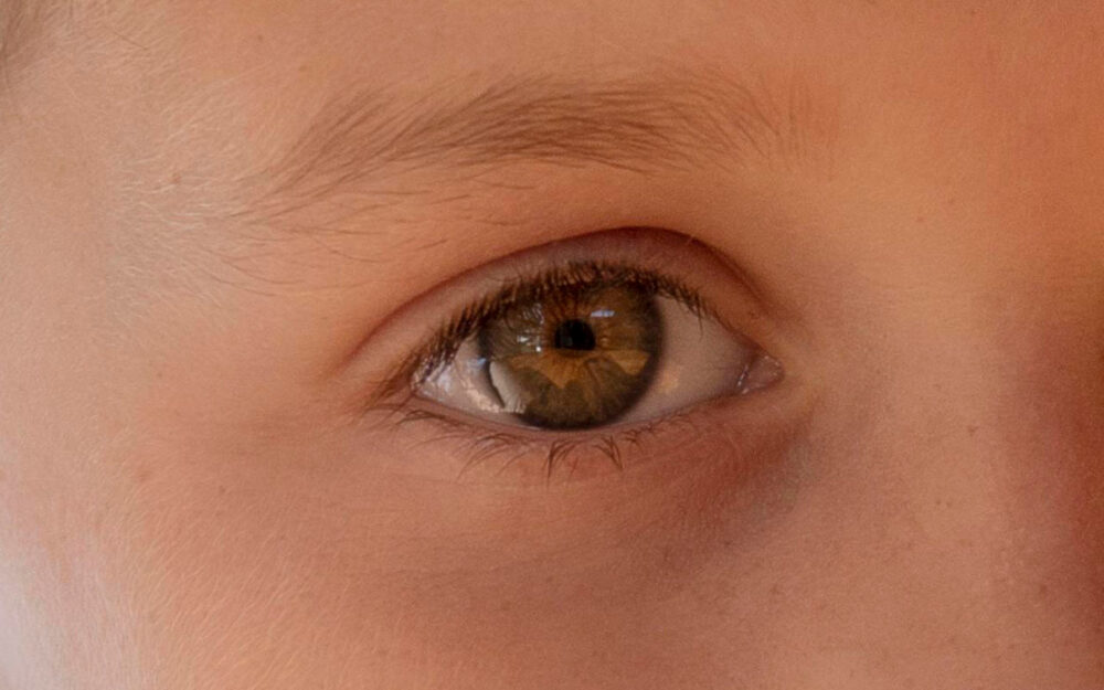
AI Diagnoses Autism with 100% Accuracy Using Retinal Images
Researchers from Yonsei University College of Medicine in South Korea have developed a method to diagnose autism and assess its severity in children using photographs of the retina. This approach utilizes a deep learning artificial intelligence algorithm, which demonstrated 100% accuracy in diagnosing autism.
The retina, which connects to the optic nerve, is part of the central nervous system and serves as a window to the brain. This makes it possible to easily and non-invasively gather important information about the brain. Previously, scientists in the UK created a rapid concussion diagnosis method using a safe retinal laser. Now, Korean researchers have introduced a new technique.
Study Details and Methodology
The study involved 958 children with an average age of 7-8 years, resulting in 1,890 retinal images. Half of the participants were diagnosed with autism spectrum disorder (ASD), while the other half formed a control group matched by age and gender. The severity of ASD symptoms was assessed using the ADOS-2 and SRS-2 scales.
A convolutional neural network, a deep learning algorithm, was trained on 85% of the images and symptom severity scores to build models for ASD screening and symptom assessment. The remaining 15% of images were used for testing.
Results: Unprecedented Accuracy
For ASD screening on the test set, the AI was able to identify children with ASD with an average area under the receiver operating characteristic curve (AUROC) of 1.00, indicating 100% prediction accuracy in this experiment. The AUROC value ranges from 0 to 1. Notably, there was no significant decrease in the average AUROC even when 95% of the least important image areas, excluding the optic disc, were removed.
The researchers noted that the models showed promising results in distinguishing ASD from typical development in children using retinal photographs. The optic disc area proved especially important. Interestingly, the models maintained an average AUROC of 1.00 using only 10% of the image containing the optic disc, highlighting this region’s critical role in differentiating ASD from typical development.
The average AUROC for assessing symptom severity was 0.74, which is considered “acceptable” (0.8 to 0.9 is “excellent”). The scientists stated that retinal photographs could provide additional information about the severity of symptoms.
Implications and Future Research
Study participants were aged four and older. Based on the results, the researchers suggest their AI model could be used as a screening tool starting at this age. Since a newborn’s retina continues to develop until age four, further research is needed to determine the tool’s accuracy in younger children.
The researchers emphasized that while further studies are required to confirm the generalizability of the results, their work is a significant step toward developing objective ASD screening tools. This could help address urgent issues such as limited access to specialized psychiatric assessments for children due to resource constraints.


